Augmented Reality Guided Surge: Next Generation Fluorescent and Image Guided Surgery

Reduce the time and cost while improving patient outcomes by revolutionizing visualizations during surgery using augmented reality
Arlington, VA United States Medical Device Connected Health
About our project
The problem we solve: Other than infection, tissue differentiation is the biggest surgical challenge – where, what and how much to cut. Verbal and visual distractions from 2D monitors lengthen procedures and negatively impact patient outcomes. Current fluorescent guided surgery (FGS) approaches have several major shortcomings: 1) cost; 2) usability; 3) complexity; and 4) low adoption rates. Current FGS solutions are expensive ($1.3M to $2.3M more expensive over 3 years based on 20 surgeries per week and 40 surgical weeks). Current FGS solutions have several usability concerns: black and white, 2D images outside of the surgical field, single focal point, requirement to turn off lights in order to obtain fluorescence, bulky hardy requires extra time (15-30 minutes per procedure) to set up and mechanical arm for camera and light sources are hard to maneuver. The use of external monitors.

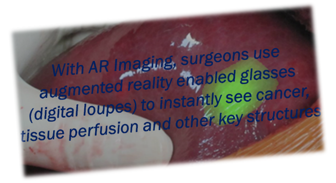
About our solution: With AR Imaging, a surgeon puts on AR-enabled glasses (digital loupes) with variable magnification (2x-8x), adjustable color contrasts and voice navigation. Vivid 3D color images of the surgical field and fluorescent imaging significantly improve tissue differentiation and depth perception. Preoperative diagnostic images (e.g. CT, MRI) superimposed on the patient improve situational awareness of the surrounding structures and organs. By revolutionizing clinician visualizations and situational awareness, AR Imaging is focused on eliminating verbal and visual distractions and improving decision making to reduces the time and cost while improving patient outcomes from medical and surgical procedures.

Progress to date:
|
About Our Team

Creator: Michael Riemer
Location: Virginia
Bio: I am a technology business executive and "accidental" serial entrepreneur who left medical school to start (and exit) my first company. 32 years of B2B enterprise software leadership experience in product management, marketing and business development including 3 exits. Last 10 years leading Industrial IoT and field service applications, platforms and digital ecosystems for connected vehicles and heavy equipment Early executive at Nextel (1993). 7+ years running products (handset, services, data, applications) including creating/launching GTM segment strategy (7 segments, 25M Pops) that delivered $70 ARPU (industry highest), 2.5% churn (industry lowest), 4.5M subscribers (record growth) and 3500% increase in market cap. Unique combination of software, hardware and telecom product management with more than 20 product launches and 5 granted patents Broad industry experience (financial services, heavy equipment, transportation, cybersecurity, telecom, field service). Extensive technology background (IoT, hardware, software, cloud, mobile, voice recognition, AI/ML, Predictive & Prescriptive Analytics) generating >$200M in software contract value.
Title: Chief Executive Officer
About Team Members
Frank Papay
Chief Medical Officer, MD, MS BME, FACS, FAAP
Biography: Chairman, Dermatology and Plastic Surgery Institute at the Cleveland Clinic. Performed first near-total face transplant in US and commercialized first neuromodulation implant device.
Title: Chief Medical Officer
Advanced Degree(s): MD, MS BME, FACS, FAAP
Twitter:
@4arimaging
LinkedIn:
https://www.linkedin.com/in/frank-a-papay-md-3193187/
Yang Liu
CTO, PhD Biomedical Engineering
Biography: Assistant Professor, Biomedical Engineering, University of Akron
Expert in medical virtual reality and intraoperative imaging; Principal investigator of multiple research grants from US Air Force, Army, NASA, ODOD, etc; Young Investigator award recipient from International Society of Computer Aided Surgery.
Title: CTO
Advanced Degree(s): PhD Biomedical Engineering
Twitter:
@4arimaging
LinkedIn:
https://www.linkedin.com/in/-yangliu/
Andrew Moan
CPO, MBA, Finance
Biography: General Manager, Cleveland Clinic – Digital Innovations (AR / VR / AI)
Expert in product development for AR / VR / AI. 15+ years in hi-tech focusing on simulation, computer hardware development, software design and business evaluation. Previous employment at Microsoft, Apple, and McKinsey & Co.
Title: CPO
Advanced Degree(s): MBA, Finance
Twitter:
@4arimaging
LinkedIn:
https://www.linkedin.com/in/andrewmoan/
About Our Company

AR Imaging
Location: 4650 Washington Blvd
Apt 118
Arlington, Virginia 22201
Founded: 2015
Website: https://www.arimaging.com
Blog: https://www.arimaging.com/blogs/
Twitter: @4arimaging
Facebook: https://www.facebook.com/4arimaging/
Product Stage: Prototype/MVP
YTD Sales: Working on it
Employees: 3-5
How We Help Physicians
|
AR Imaging uses augmented reality to revolutionize clinical visualizations to enhance tissue differentiation. We do this by providing AR-enabled glasses (digital loupes), that enable the surgeon to view 3D color images of the patient with coregistration of fluorescent contrast agents and pre-operative diagnostic images (e.g. CT, MRI) superimposed on the surgical field. Fluorescent imaging helps quickly identify tissue perfusion (viability) and helps identify cancerous areas (they light up like a Christmas tree). The superimposed preoperative images provide a highly effective image guided surgery solution that improves decision making and situational awareness. All targeted procedures have current solutions and related DRGs and ICDs for reimbursements and payments associated with fluorescent and image guided surgery. In addition to providing a major cost savings, these solutions have other shortcomings that impact surgical experiences. This includes:
Specifically, the following value for each user that addresses their respective pain point(s) is listed in the table below:
|
How We Help Hospitals
|
Hospital |
|
|
Innovation Details
Intellectual Property Summary
|
3 PCT patent applications, the first of which has had office action and several key claims already approved. AR Imaging has the exclusive WW license from Univ. of Akron.
|
Clinical Information
Animal studies completed and published.
A miniature wearable optical imaging system for guiding surgeries
Image guidance can result in improved surgical outcomes, shorter operating times as well as a reduced likelihood of requiring a follow-up surgery for various medical interventions. Many intraoperative imaging systems utilize 2D computer monitors, making it difficult to correlate the surgical landscape with the displayed functional information as well as potentially distracting the surgeon. To address this issue, a miniature, wearable Near Infrared (NIR) fluorescent imaging system entitled Stereoscopic Optical Imaging Goggle is developed. The system is made up of two imaging sensors affixed to a wearable stereoscopic display, providing the surgeon with functional data in 3 dimensions with depth perception. We have characterized the system’s optical properties and fluorescent detection limits. In addition, we have demonstrated the efficacy of the system during surgical studies in chicken. We have found that the system can resolve fluorescent structures down to 0.25mm. The system was successfully guided the excision of fluorescent tissue from a chicken. To the best of our knowledge, the Stereoscopic Optical Imaging Goggle is the first wearable wide-field fluorescence imaging system that offers stereoscopic imaging capability and 3D depth perception.
Stereoscopic Integrated Imaging Goggles for Multimodal Intraoperative Image Guidance
We have developed novel stereoscopic wearable multimodal intraoperative imaging and display systems entitled Integrated Imaging Goggles for guiding surgeries. The prototype systems offer real time stereoscopic fluorescence imaging and color reflectance imaging capacity, along with in vivo handheld microscopy and ultrasound imaging. With the Integrated Imaging Goggle, both wide-field fluorescence imaging and in vivo microscopy are provided. The real time ultrasound images can also be presented in the goggle display. Furthermore, real time goggle-to-goggle stereoscopic video sharing is demonstrated, which can greatly facilitate telemedicine. In this paper, the prototype systems are described, characterized and tested in surgeries in biological tissues ex vivo. We have found that the system can detect fluorescent targets with as low as 60 nM indocyanine green and can resolve structures down to 0.25 mm with large FOV stereoscopic imaging. The system has successfully guided simulated cancer surgeries in chicken. The Integrated Imaging Goggle is novel in 4 aspects: it is (a) the first wearable stereoscopic wide-field intraoperative fluorescence imaging and display system, (b) the first wearable system offering both large FOV and microscopic imaging simultaneously, (c) the first wearable system that offers both ultrasound imaging and fluorescence imaging capacities, and (d) the first demonstration of goggle-to-goggle communication to share stereoscopic views for medical guidance.
Fluorescence Imaging Topography Scanning System for intraoperative multimodal imaging
Fluorescence imaging is a powerful technique with diverse applications in intraoperative settings. Visualization of three dimensional (3D) structures and depth assessment of lesions, however, are oftentimes limited in planar fluorescence imaging systems. In this study, a novel Fluorescence Imaging Topography Scanning (FITS) system has been developed, which offers color reflectance imaging, fluorescence imaging and surface topography scanning capabilities. The system is compact and portable, and thus suitable for deployment in the operating room without disturbing the surgical flow. For system performance, parameters including near infrared fluorescence detection limit, contrast transfer functions and topography depth resolution were characterized. The developed system was tested in chicken tissues ex vivo with simulated tumors for intraoperative imaging. We subsequently conducted in vivo multimodal imaging of sentinel lymph nodes in mice using FITS and PET/CT. The PET/CT/optical multimodal images were co-registered and conveniently presented to users to guide surgeries. Our results show that the developed system can facilitate multimodal intraoperative imaging.
|
Intraoperative Fluorescence Imaging and Multimodal Surgical Navigation Using Goggle System Intraoperative imaging is an invaluable tool in many surgical procedures. We have developed a wearable stereoscopic imaging and display system entitled Integrated Imaging Goggle, which can provide real-time multimodal image guidance. With the Integrated Imaging Goggle, wide field-of-view fluorescence imaging is tracked and registered with intraoperative ultrasound imaging and preoperative tomography-based surgical navigation, to provide integrated multimodal imaging capabilities in real-time. Herein we describe the system instrumentation and the methods of using the Integrated Imaging Goggle to guide surgeries. |
AR Imaging has a working prototype being used in surgery in trials at the Cleveland Clinic. Initial surgery performed by Dr. Stephen R. Grobmyer, Director, Surgical Oncology at the Cleveland Clinic.
The first active IRB is directed toward sentinel node lymph node biopsies for breast cancer (NCT02802553), the second concerns basal/squamous cell carcinoma (skin cancers) auto-fluorescence with Levulon (alpha-levulinic acid) versus actinic keratosis differentiation and the third submitted IRB is perfusion of reconstruction flaps in plastic surgery mainly free flaps in breast reconstruction. Approved IRBs at CCF go through a lengthy process that also includes Case Western Reserve Medical School, University Hospitals and Metro General)
Regulatory Status
Our device is type 2 and will require a 510K. However there are several strong predicates and we have a pre-submission document almost completed
How we will use the funds raised
|
$1.5M - MVP for fluorescent guided surgery. Finalize the digital loupe design and manufacturer the initial MVP. It will also be used to complete the current trials and obtain FDA and EU approvals.
$2.0M - Version 1 launch and support/success teams in place. Post market studies with KOLs. R&D on new tissue and bone/organ visualizations as well software and accessories. Customer success and support teams.
|
Thank You
(This post can be read more easily here)
Return on Heartbeat: The Importance of Being Mission Driven
Why did you join AR Imaging is a frequent question these days. The answer is both simple and personal.
After 32 years as a serial entrepreneur, I was looking for something more meaningful. Helping deliver $100,000,000s of software and technology had added little to society.
AR Imaging is also in my hometown, Cleveland, Ohio. (No Browns jokes please.) And, I want to contribute to its blossoming technology and entrepreneurial ecosystem.
It's also exciting to contribute to a solution that can help deliver faster, better and less expensive cancer surgeries. This is near and dear to me because my mom died of cancer at 45.
Finally, I wanted a new opportunity to create mission-driven values and culture. One that makes people as critical as the product and customer. An opportunity to create measurable value for employees as well as the world around us.
I call this Return on Heartbeat.
My Journey
After 32 years of early and growth stage ventures, a medical device venture may seem like a big change.
Yet, my career has crossed many different technologies , applications and industries.
Growing up on the eastside of Cleveland, I attended Beachwood High School. After, I went "pre-med" to the University of Rochester where I majored in Biology and Philosophy.
After graduation, Case Western Reserve University, School of Medicine extended an offer. Shortly after accepting, Peter Tippett and I started a software company, Certus International. It was one of the first anti-virus products for PCs. The company was eventually sold to Symantec.
Since then, Peter and I have both enjoyed great careers but never found another opportunity to work together, until now.
The Company
In early February 2018, Peter suggested that I look into a company which was then called I-Imaging. A few weeks later we traveled to Cleveland. I met the co-founders Dr. Frank Papay and Yang Liu, PhD. A month later, I was onboard and very excited.
AR Imaging combines many great technologies. This includes augmented reality, digital health and industrial IoT. It also uses techniques in computer vision as well as image recognition and processing.
But technologies don't solve problems.
So let's not focus on them for now.
Except to credit AR Imaging's tremendous technical achievements.
Tissue Differentiation Is The Focus
Instead, let's focus on a critical problem - tissue differentiation.
Besides infection, knowing what, where and how much to cut is a huge challenge. It is one of the oldest and most basic challenges of surgery. It takes year of training and practice to ensure successful outcomes.
AR Imaging's goal is to improve decision-making by improving clinical visualizations of tissue differentiation. We also happen to be using a bunch of "cool" technologies.
AR Imaging is the next generation of fluorescent and image guide surgery. Improving decision-making and situational awareness. Our objective is to improve patient outcomes and lessen the cost and time of the procedures.
We are very optimistic of our commercial success but what about return on heartbeat?
World Mission View
By 2050, the US will see an 82% growth of individuals over the age 65. China will experience 183% in that same timeframe. Europe’s aging projections are similar. Our aging population is increasing the frequency of related procedures including cancer.
At the same time, the WHO estimates a shortage of 4.3 million physicians, nurses and other health workers. In the US, the AMA estimates a shortage of 91,500 physicians by 2020 and up to 130,600 by the year 2025.
Contributing to 5 billion people today without access to safe surgery.
AR Imaging is not a panacea to this enormous problem.
But AR Imaging can help delivery safe surgery solutions around the globe.
Our small form factor and cost effective approach provides a great delivery platform. First for fluorescent and image guided surgery. In the future for telehealth, telesurgery and other collaborative solutions.
The Circle of Life
My journey has led me back to my hometown. To the medical industry. To a mission driven opportunity. To a great return on heartbeat.
Supporters
Index Score
7
Score
0
Score
-
This campaign has ended but you can still get involved.See options below.
Help us find best new ideas to fund by telling us what you think. Your feedback goes straight to the team behind this project in private, so tell them what you really think.
Important Disclosure: MedStartr.com is a website owned and operated by MedStartr, Inc., which is not a broker-dealer, funding portal or investment advisor; and neither the website nor MedStartr, Inc. participate in the offer or sale of securities. All securities related activity is conducted through Young America Capital, LLC, a registered broker-dealer and member of FINRA/SIPC. No communication, through this website, email or in any other medium, should be construed as a recommendation for any securities offering.

